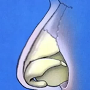
A pollybeak deformity refers to an excess prominence, or contour, in the region of the nose immediately above the tip of the nose. A pollybeak is typically classified as being a result of either cartilage or scar. Therefore, you will often see the terms cartilaginous pollybeak and soft tissue (scar) pollybeak used, respectively, when discussing this rhinoplasty topic. In most cases, a pollybeak deformity results from prior rhinoplasty surgery. It is much less common to see a pollybeak deformity that is congenital in nature.
Cartilaginous Pollybeak Deformity Following Prior Rhinoplasty
A cartilaginous pollybeak deformity usually arises in someone who had prior rhinoplasty to reduce the bridge height. Normally these are rhinoplasty patients who originally had a dorsal hump, or bump. In most cases of a dorsal hump, reduction of the bridge involves taking down both the cartilage and the bone to achieve a nice smooth, nasal profile. Unfortunately, some rhinoplasty surgeons fail to adequately remove the entire cartilage portion of the bump. This exact point is demonstrated visually in the adjacent rhinoplasty diagrams.

On the left is a nose with a dorsal hump or bump. When you remove a hump like this, you need to reduce the height of the cartilage (yellow) and bone (blue) in a fairly equivalent fashion to achieve a nice straight, smooth profile. The middle photo below shows what the ideal goal of hump reduction is in rhinoplasty. In a pollybeak deformity resulting from prior rhinoplasty, some of the cartilage that should have been removed is still present. This area of residual cartilage is referred to as the supratip region. As is seen below right and indicated by the white arrows with red outline, the lower portion of the bridge cartilage is still intact while the remainder of the bridge (above the red arrow) has been taken down. In this particular situation, the middle and upper bridge is at the correct height and has been reduced properly. But the supratip portion immediately above the tip region is still excessively high. This remaining portion that should have been removed along with the rest of the bridge directly accounts for the cartilaginous pollybeak deformity.
